Source: Wikipedia and some old Wk. 2 lecture
Definition:
Blood Clotting Cascade: How blood clots (The coagulation process in humans is most extensively researched and therefore best known - and therefore most complicated).
Blood clotting, or coagulation, is an important part of homeostasis (the cessation of blood loss from a damaged vessel) whereby a damaged blood vessel wall is covered by a fibrin clot to stop hemorrhage and aid repair of the damaged vessel. Disorders in coagulation can lead to increased hemorrhage and/or thrombosis and embolism.
In Brief:
The blood clotting cascade consists of a large number of factors, most of which are specialized enzymes called proteases (enzymes that cleave peptide bonds between amino acids of proteins).
It is called a "cascade" because each factor activates the next factor, usually by cleaving it.
Overall, the effect is to activate the enzyme, thrombin, which is responsible for most of the final, visible effects of clotting.
Two major functions of thrombin are the formation of a fibrin clot and the activation of platelets - together these components form a blood clot.
Thrombin:
Thrombin is a coagulation protein that has many effects in the coagulation cascade. It is a serine protease that converts soluble fibrinogen into insoluble strands of fibrin, as well as catalyzing many other coagulation-related reactions.
Thrombosis:
Thrombosis is the formation of a clot, or thrombus, inside a blood vessel - obstructing blood flow through the circulation system.
If the clotting cascade is activated inappropriately or in an uncontrolled manner, a thrombosis is formed. This is usually due to a narrowing of an artery or vein.
Venouse thrombosis (deep vein thrombosis - DVT) is usually due to lack of control by anticoagulant factors.
Antithrombin:
(a serine protease inhibitor [serpine] under conformational control)
Antithrombin is a small molecule that inactivates several enzymes of the coagulation system. It is a glycoprotein produced by the liver.
Antithrombin inhibits the serine proteases of the clotting cascade, especially thrombin and factor Xa and thus plays a fundamental role in controlling the blood coagulation cascade.
Serpins:
Lecture: "Serpins - a family of proteinase inhibitors that undergo conformational change"
Wikipedia: Serpins (short for serine protease inhibitor) are a group of structurally related proteins, many of which inhibit peptidases (enzymes that degrade protein, old name: proteases). Although initially simply considered a class of protease inhibitors (proteins that block the action of peptidases), it was discovered later that it has members that do not inhibit any enzymes, but serve as storage proteins (ovalbumin, in egg white), carriage proteins (thyroxine-binding globulin, sex hormone binding globulin) and hormone precursors (angiotensinogen). The term serpin is used for these members as well, despite their noninhibitory function.
Heparin:
Heparin is a highly sulfated glycosaminoglycan widely used as an injectable anticoagulant. It is also used to form an inner anticoagulant surface on various experimental and medical devices such as test tubes and renal dialysis machines. Pharmaceutical grade heparin is commonly derived from mucosal tissues of slaughtered meat animals such as porcine intestine or bovine lung.
Heparin controls the action of antithrombin. For efficient interaction with target proteinases antithrombin requires the cofactor heparin:
- Antithrombin circulates in a relatively inactive form
- Binding heparin dramatically accelerates interaction with target proteinases
- Major anticoagulant effect of heparin occurs by acting through antithrombin
Mutations in antithrombin - a major risk factor for DVT
- Mice completely deficient in antithrombin die in utero due to uncontrolled clotting - antithrombin is important
- Heterozygous mutations in antithrombin (mutation to one allele) can cause it to be ineffective in controlling clotting proteases
- Lack of efficient control of the clotting proteases can lead to venous thrombosis
Causes of antithrombin deficiency:
- Antithrombin, like other serpins, is prone to being unstable
- Certain mutations cna cause it to be unstable because it aggregates or polymerises
- This means there will be lower levels of active antithrombin
- Point mutations to antithrombin may also cause it to be inactive without affecting stability
- These mutations usually affect residues important for binding heparin or the protease target
Showing posts with label Biochemistry and Molecular Biology. Show all posts
Showing posts with label Biochemistry and Molecular Biology. Show all posts
1/8/07
Myoglobin
Source: Wikipedia and some random lecture I once had
Definition:
Myoglobin is a protein that consists of 153 amino acids and a heme (iron containing porphyrim) prosthetic group
Importance:
Myoglobin is the primary oxygen-carrying pigment of muscle tissues (found in muscles, stores oxygen).
The structure of the myoglobin protein is specifically adapted to provide a pocket to bind the heme group.
Role in Disease:
Myoglobin is released from damaged muscle tissue (rhabdomyolysis), which has very high concentrations of myoglobin. The released myoglobin is filtered by the kidneys but is toxic to the renal tubular epithelium and so may cause acute renal failure.
Myoglobin is a sensitive marker for muscle injury, making it a potential marker for heart attack in patients with chest pain. Its lack of specificity and the cost of the analysis has prevented its widespread use.
Definition:
Myoglobin is a protein that consists of 153 amino acids and a heme (iron containing porphyrim) prosthetic group
Importance:
Myoglobin is the primary oxygen-carrying pigment of muscle tissues (found in muscles, stores oxygen).
The structure of the myoglobin protein is specifically adapted to provide a pocket to bind the heme group.
Role in Disease:
Myoglobin is released from damaged muscle tissue (rhabdomyolysis), which has very high concentrations of myoglobin. The released myoglobin is filtered by the kidneys but is toxic to the renal tubular epithelium and so may cause acute renal failure.
Myoglobin is a sensitive marker for muscle injury, making it a potential marker for heart attack in patients with chest pain. Its lack of specificity and the cost of the analysis has prevented its widespread use.
Amino Acids
"Golly gosh" ... so proteins are complex biological compounds made of amino acids,
... and amino acids are like molecules that have amine (groups that have Nitrogen as key atom) and carboxyl (groups characterized by the presence of carboxyl: -COOH) functioning groups. ?? So is N-COOH an amino acid? And three nucleotides arrange each amino acid?? GaH! Must... consult... wikipedia...
Amino Acids
In chemistry, an amino acid is any molecule that contains both amine and carboxyl functional groups. In biochemistry, this term is used to refer to alpha amino acids: molecules where the amino and carboxylate groups are attached to the same carbon, the α–carbon.
These alpha amino acids are the basic components of proteins. There are twenty standard amino acids used by cells in protein biosynthesis and these are specified by the general genetic code. These twenty amino acids can be biosynthesised from simpler molecules, but organisms differ in how many they are able to produce and essential amino acids must be obtained in their diet.
In proteins, an amino acid residue is what is left of an amino acid once a molecule of water has been lost (an H+ from the nitrogenous side and an OH- from the carboxylic side) in the formation of a peptide bond. These are the chemical bonds that links the amino acid monomers into a protein chain. Each different protein has a unique amino acid sequence that is known as its primary structure. Just as the letters of the alphabet can be combined in different ways to form an almost endless variety of words, amino acids can be linked together in varying sequences to form a huge variety of proteins. Each unique sequence of amino acids folds up to form a unique three-dimensional structure, the protein's tertiary structure. These tertiary structures determine the functions of proteins.
Amino acids are the basic structural building units of proteins. They form short polymer chains called peptides or longer chains either called polypeptides or proteins. The process of such formation from an mRNA template is known as translation, which is part of protein synthesis.
Some of the 20 standard proteinogenic amino acids are called essential amino acids because the human body cannot synthesize them from other compounds through chemical reactions, and they therefore must be obtained from food. Histidine and arginine are generally only considered essential in children, because the metabolic pathways that synthesize these amino acids are not fully developed in children. Also some amino acid supplements are said to be dangerous by dietitians since the human body needs a certain specific balance. So taking some single amino acid supplements will throw off this balance and can cause health problems. For further information please read Protein in nutrition.
... and amino acids are like molecules that have amine (groups that have Nitrogen as key atom) and carboxyl (groups characterized by the presence of carboxyl: -COOH) functioning groups. ?? So is N-COOH an amino acid? And three nucleotides arrange each amino acid?? GaH! Must... consult... wikipedia...
Amino Acids
In chemistry, an amino acid is any molecule that contains both amine and carboxyl functional groups. In biochemistry, this term is used to refer to alpha amino acids: molecules where the amino and carboxylate groups are attached to the same carbon, the α–carbon.
These alpha amino acids are the basic components of proteins. There are twenty standard amino acids used by cells in protein biosynthesis and these are specified by the general genetic code. These twenty amino acids can be biosynthesised from simpler molecules, but organisms differ in how many they are able to produce and essential amino acids must be obtained in their diet.
In proteins, an amino acid residue is what is left of an amino acid once a molecule of water has been lost (an H+ from the nitrogenous side and an OH- from the carboxylic side) in the formation of a peptide bond. These are the chemical bonds that links the amino acid monomers into a protein chain. Each different protein has a unique amino acid sequence that is known as its primary structure. Just as the letters of the alphabet can be combined in different ways to form an almost endless variety of words, amino acids can be linked together in varying sequences to form a huge variety of proteins. Each unique sequence of amino acids folds up to form a unique three-dimensional structure, the protein's tertiary structure. These tertiary structures determine the functions of proteins.
Amino acids are the basic structural building units of proteins. They form short polymer chains called peptides or longer chains either called polypeptides or proteins. The process of such formation from an mRNA template is known as translation, which is part of protein synthesis.
Some of the 20 standard proteinogenic amino acids are called essential amino acids because the human body cannot synthesize them from other compounds through chemical reactions, and they therefore must be obtained from food. Histidine and arginine are generally only considered essential in children, because the metabolic pathways that synthesize these amino acids are not fully developed in children. Also some amino acid supplements are said to be dangerous by dietitians since the human body needs a certain specific balance. So taking some single amino acid supplements will throw off this balance and can cause health problems. For further information please read Protein in nutrition.
1/7/07
Protein
Source: Wikipedia
Definition:
Protein: large organic compounds made of amino acids.
History:
The name protein comes from the Greek πρώτα ("prota"), meaning "of primary importance" and were first described and named by Jöns Jakob Berzelius in 1838. However, their central role in living organisms was not fully appreciated until 1926, when James B. Sumner showed that the enzyme urease was a protein. The first protein structures to be solved included insulin and myoglobin; the first was by Sir Frederick Sanger who won a 1958 Nobel Prize for it, and the second by Max Perutz and Sir John Cowdery Kendrew in 1958. Both proteins' three-dimensional structures were amongst the first determined by x-ray diffraction analysis; the myoglobin structure won the Nobel Prize in Chemistry for its discoverers.
Definition:
Protein: large organic compounds made of amino acids.
History:
The name protein comes from the Greek πρώτα ("prota"), meaning "of primary importance" and were first described and named by Jöns Jakob Berzelius in 1838. However, their central role in living organisms was not fully appreciated until 1926, when James B. Sumner showed that the enzyme urease was a protein. The first protein structures to be solved included insulin and myoglobin; the first was by Sir Frederick Sanger who won a 1958 Nobel Prize for it, and the second by Max Perutz and Sir John Cowdery Kendrew in 1958. Both proteins' three-dimensional structures were amongst the first determined by x-ray diffraction analysis; the myoglobin structure won the Nobel Prize in Chemistry for its discoverers.
Biochemistry:
Proteins are large organic compounds made of amino acids via peptide bonds (amino acids share carboxyl atom of on group and the amine of another). The sequence of amino acids defines the protein, and the sequence is arranged by a certain gene in DNA. There are 20 "standard" amino acids coded by human DNA. Different proteins can work together to carry out different functions.
Once linked in the protein chain, an individual amino acid is called a residue and the linked series of carbon, nitrogen, and oxygen atoms are known as the main chain or protein backbone.
There is some ambiguity between the usage of the words protein, polypeptide, and peptide. Protein is generally used to refer to the complete biological molecule in a stable conformation, while peptide is generally reserved for a short amino acid oligomers often lacking a stable 3-dimensional structure. However, the boundary between the two is ill-defined and usually lies near 20-30 residues. Polypeptide can refer to any single linear chain of amino acids, usually regardless of length, but often implies an absence of a single defined conformation.
Synthesis:
Proteins are assembled from amino acids using information encoded in genes.
(Amino acids are molecules that contain both amine and carboxyl-funtional groups (alpha amino acids). There are 20 standard amino acids used by cells in protein biosynthesis and these are specified by the general genetic code.)
(Amino acids are molecules that contain both amine and carboxyl-funtional groups (alpha amino acids). There are 20 standard amino acids used by cells in protein biosynthesis and these are specified by the general genetic code.)
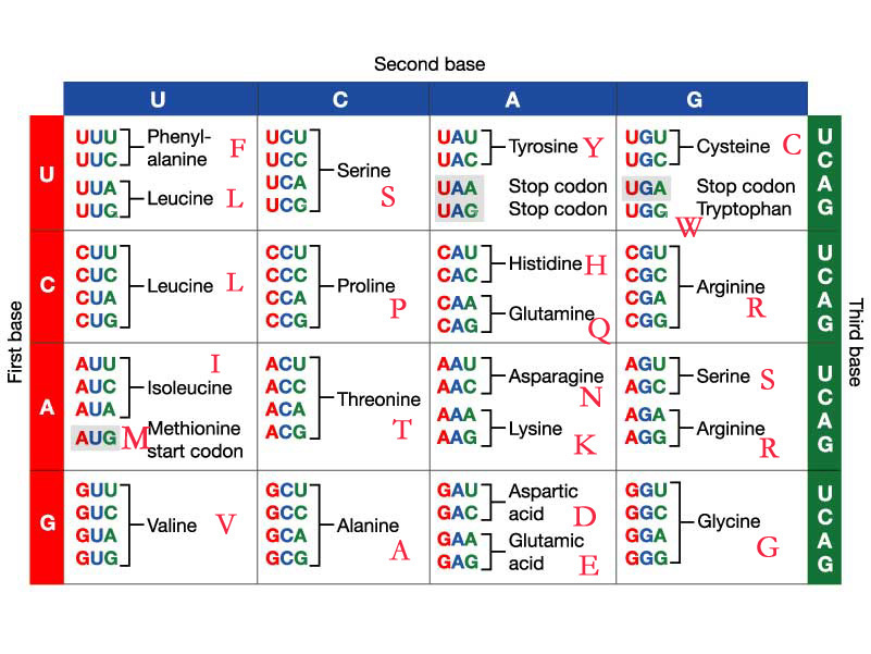
Each protein has its own unique amino acid sequence that is specified by the nucleotide sequence of the gene encoding this protein. The genetic code is a set of three-nucleotide sets called codons and each three-nucleotide combination stands for an amino acid, for example ATG stands for methionine. Because DNA contains four nucleotides, the total number of possible codons is 64; hence, there is some redundancy in the genetic code and some amino acids are specified by more than one codon.

Structure:
Proteins contain 4 levels of structure:
1). Primary: the amino acid sequence
2). Secondary: regularly repeating structures shaped by hydrogen bonds (most common are the alpha helix and the beta sheet)
3). Tertiary: the overall shape of a single protein molecule; the term "tertiary structure" is often used as synonymous with the term fold. Tertiary structured proteins are polypeptide chains consisting of secondary structured protein twisted and folded into a more 3-D structure.
4). Quaternary: contains a number of tertiary structured proteins as sub-units, which function as part of the larger assembly, or protein complex.
1/6/07
Erythrocytes (Red Blood Cells)
Red Blood Cells
Source: Wikipedia
Also known as erythrocytes (from Greek erythros for "red" and kytos for "hollow", with cyte nowaday translated as "cell"). Primary function of transporting gases and other metabolites. Principle gases carried: oxygen (lungs to tissues), and carbon dioxide (tissues to lungs).
Erythrocytes consist mainly of hemoglobin, which is a conplex molecule containing heme groups (which contain iron atoms at their center - thus making blood appear red). The iron component of hemoglobin temporarily link to oxygen molecules in the lungs or gills. Oxygen can easily diffuse through the red blood cell's membrane. Hemoglobin also carries Carbox Dioxide (return waste product) back from the tissues. In mammals, erythrocytes are anucleate which means that they do not have a cell nucleus, and this no DNA (in comparison, all other vertebrates have nuclei, except for salamanders). Erythrocytes do not have many other organelles either, and thus lack the ability of repair giving them very small life spans.
The diameter of the typical erythrocyte is 6-8 µm (smaller than most other cells). A typical erythrocyte contains about 270 million hemoglobin molecules, which each contain 4 heme groups.
Women have 4-5 million erythrocytes per cubic milliliter (micrometer), and men have about 5-6 million. Collectively, red blood cells store 3.5 grams of iron.
The process by which red blood cells are made is called erythropoeisis. Erythrocytes are continuously being produced in the red bone marrow of large bones, at a rate of about 2 million per second. (In the embryo, the liver is the main site of red blood cell production.) The production can be stimulated by the hormone erythropoietin (EPO), which is used for doping in sports (because more erythrocytes = more capacity to store oxygen).
After leaving the bone marrow, they are known as reticulocytes which comprise about 1% of circulating red blood cells (they are called reticulocytes because of the reticular, or mesh-like, network of ribosomal RNA visible when stained). The number of reticulocytes is a good indicator of bone marrow activity, because it represents recent production. This means that the reticulocyte count, and the reticulocyte production index that can be calculated from it, can be used to determine whether a production problem is contributing to the anaemia, and can also be used to monitor the progress of treatment for anaemia.
Erythrocytes develop from stem cells through reticuloctyes to mature erythrocytes in about 7 days and live a total of about 120 days. The aging cells swell up to a sphere-like shape and are engulfed by phagocytes, destroyed and their materials are released into the blood. The main sites of destruction are the liver and the spleen. The heme constituent of hemoglobin is eventually excreted as bilirubin.
The blood types of humans are due to variations in surface glycoproteins of erythrocytes.
Red blood cells can be separated from blood plasma by centrifugation. During plasma donation, the red blood cells are pumped back into the body right away, and the plasma is collected.
Other than taking EPO, some athletes have tried to improve their performance by doping their blood: First about 1 liter of their blood is extracted, then the red blood cells are isolated, frozen and stored, to be reinjected shortly before the competition. (Red blood cells can be conserved for 5 weeks at −78 °C.) This practice is hard to detect but may endanger the human cardiovascular system which is not equipped to deal with blood of the resulting higher viscosity.
Importance in disease:
Sickle Cell Anaemia
Source: Wikipedia
Also known as erythrocytes (from Greek erythros for "red" and kytos for "hollow", with cyte nowaday translated as "cell"). Primary function of transporting gases and other metabolites. Principle gases carried: oxygen (lungs to tissues), and carbon dioxide (tissues to lungs).
Erythrocytes consist mainly of hemoglobin, which is a conplex molecule containing heme groups (which contain iron atoms at their center - thus making blood appear red). The iron component of hemoglobin temporarily link to oxygen molecules in the lungs or gills. Oxygen can easily diffuse through the red blood cell's membrane. Hemoglobin also carries Carbox Dioxide (return waste product) back from the tissues. In mammals, erythrocytes are anucleate which means that they do not have a cell nucleus, and this no DNA (in comparison, all other vertebrates have nuclei, except for salamanders). Erythrocytes do not have many other organelles either, and thus lack the ability of repair giving them very small life spans.
The diameter of the typical erythrocyte is 6-8 µm (smaller than most other cells). A typical erythrocyte contains about 270 million hemoglobin molecules, which each contain 4 heme groups.
Women have 4-5 million erythrocytes per cubic milliliter (micrometer), and men have about 5-6 million. Collectively, red blood cells store 3.5 grams of iron.
The process by which red blood cells are made is called erythropoeisis. Erythrocytes are continuously being produced in the red bone marrow of large bones, at a rate of about 2 million per second. (In the embryo, the liver is the main site of red blood cell production.) The production can be stimulated by the hormone erythropoietin (EPO), which is used for doping in sports (because more erythrocytes = more capacity to store oxygen).
After leaving the bone marrow, they are known as reticulocytes which comprise about 1% of circulating red blood cells (they are called reticulocytes because of the reticular, or mesh-like, network of ribosomal RNA visible when stained). The number of reticulocytes is a good indicator of bone marrow activity, because it represents recent production. This means that the reticulocyte count, and the reticulocyte production index that can be calculated from it, can be used to determine whether a production problem is contributing to the anaemia, and can also be used to monitor the progress of treatment for anaemia.
Erythrocytes develop from stem cells through reticuloctyes to mature erythrocytes in about 7 days and live a total of about 120 days. The aging cells swell up to a sphere-like shape and are engulfed by phagocytes, destroyed and their materials are released into the blood. The main sites of destruction are the liver and the spleen. The heme constituent of hemoglobin is eventually excreted as bilirubin.
The blood types of humans are due to variations in surface glycoproteins of erythrocytes.
Red blood cells can be separated from blood plasma by centrifugation. During plasma donation, the red blood cells are pumped back into the body right away, and the plasma is collected.
Other than taking EPO, some athletes have tried to improve their performance by doping their blood: First about 1 liter of their blood is extracted, then the red blood cells are isolated, frozen and stored, to be reinjected shortly before the competition. (Red blood cells can be conserved for 5 weeks at −78 °C.) This practice is hard to detect but may endanger the human cardiovascular system which is not equipped to deal with blood of the resulting higher viscosity.
Importance in disease:
Sickle Cell Anaemia
1/2/07
Chemical Components of Cells
My next real lecture was on the chemical components of cells, a topic as exciting as it sounds. It was really just an intro lecture, aimed to revive lost high-school education.
Chemical Components of Cells
1. Water
Water's molecular structure (H2O) gives it the capacity to form hydrogen bonds (sharing of a H atom). The most important property of water is that it is an excellent solvent (can dissolve many chemical stuff, like salts, sugars, acids, and other polar molecules = hydrophillic compounds). Some stuff can't dissolve in water (hydrophobic compounds), like fats and oils and stuff. This property is important when administrating drugs.
2. pH
pH is the negative logarithm of the hydrogen ion concentration (??). Easier to note that <7>7 is basic. Buffers are somewhere in the middle (~7) and their inclusion limits a solution from changing pH easily.
3. Functional groups
I don't remember this term, but functional groups are specific groups of atoms within molecules that are responsible for the characteristic of those molecules (shape, polarity, reactivity, solubility). Examples.
4. Macromolecules
Large molecules made by smaller molecules called monomers grouping together. Macromolecules include polysaccharides, proteins and nucleic acids (they all depend on their monomers as functional groups).
5. Monomers
Monomer (from Greek mono "one" and meros "part") is a small molecule that joins other monomers via condensation reactions (when 2 compounds join and in the process lose a small molecule, usually water) to become polymers. Hydrolysis reactions (water is used to break a compound into smaller parts) break polymers into monomers.
6. Amino Acids
Amino acids are my favorites. Chemically, they are molecules that contain both amine (N-based chemicals) and carboxyl (chemicals that contain carboxyl groups, formula: -COOH) functional groups via peptide bonds (basically condensation reactions, but in amino acids the bond becomes CO-NH and is known as a peptide bond, just to confuse me). There are 20 standard amino acids which bond with each other, forming polypeptide chains of proteins in one of four 3D structures (primary, secondary, tertiary and quaternary). More on those later.
7. Monosaccharides
Monosaccharides are simple sugars (glucose, fructose). When they link up to each other via covalent bonds (sharing of one or more pairs of electrons) they become more complex: dissaccharides (maltose, lactose and sucrose), oligossaccharides and polysaccharides (starch - which stores energy in plants, and glycogen - which stores energy in animal liver and muscles).
8. Nucleic Acids
Nucleic acids are polmers made of nucleotides. Nucleotides consist of a phosphate group, a sugar (ribose in RNA and deoxyribose in DNA) and a nitrogen-containing base. Both DNA and RNA are important in forming proteins (more on this later).
9. Lipids
Lipids are often called fats (which are a subgroup of lipids called triglycerides). They are relatively insoluble in water. Phospholipids are a class of lipids that have hydrophobic hydrocarbon "tails" and hydrophillic phosphate "heads". Because the tails don't dissolve in water, they make good membranes when arranged as phospholipid bilayers.
Chemical Components of Cells
1. Water
Water's molecular structure (H2O) gives it the capacity to form hydrogen bonds (sharing of a H atom). The most important property of water is that it is an excellent solvent (can dissolve many chemical stuff, like salts, sugars, acids, and other polar molecules = hydrophillic compounds). Some stuff can't dissolve in water (hydrophobic compounds), like fats and oils and stuff. This property is important when administrating drugs.
2. pH
pH is the negative logarithm of the hydrogen ion concentration (??). Easier to note that <7>7 is basic. Buffers are somewhere in the middle (~7) and their inclusion limits a solution from changing pH easily.
3. Functional groups
I don't remember this term, but functional groups are specific groups of atoms within molecules that are responsible for the characteristic of those molecules (shape, polarity, reactivity, solubility). Examples.
4. Macromolecules
Large molecules made by smaller molecules called monomers grouping together. Macromolecules include polysaccharides, proteins and nucleic acids (they all depend on their monomers as functional groups).
5. Monomers
Monomer (from Greek mono "one" and meros "part") is a small molecule that joins other monomers via condensation reactions (when 2 compounds join and in the process lose a small molecule, usually water) to become polymers. Hydrolysis reactions (water is used to break a compound into smaller parts) break polymers into monomers.
6. Amino Acids
Amino acids are my favorites. Chemically, they are molecules that contain both amine (N-based chemicals) and carboxyl (chemicals that contain carboxyl groups, formula: -COOH) functional groups via peptide bonds (basically condensation reactions, but in amino acids the bond becomes CO-NH and is known as a peptide bond, just to confuse me). There are 20 standard amino acids which bond with each other, forming polypeptide chains of proteins in one of four 3D structures (primary, secondary, tertiary and quaternary). More on those later.
7. Monosaccharides
Monosaccharides are simple sugars (glucose, fructose). When they link up to each other via covalent bonds (sharing of one or more pairs of electrons) they become more complex: dissaccharides (maltose, lactose and sucrose), oligossaccharides and polysaccharides (starch - which stores energy in plants, and glycogen - which stores energy in animal liver and muscles).
8. Nucleic Acids
Nucleic acids are polmers made of nucleotides. Nucleotides consist of a phosphate group, a sugar (ribose in RNA and deoxyribose in DNA) and a nitrogen-containing base. Both DNA and RNA are important in forming proteins (more on this later).
9. Lipids
Lipids are often called fats (which are a subgroup of lipids called triglycerides). They are relatively insoluble in water. Phospholipids are a class of lipids that have hydrophobic hydrocarbon "tails" and hydrophillic phosphate "heads". Because the tails don't dissolve in water, they make good membranes when arranged as phospholipid bilayers.
Subscribe to:
Posts (Atom)